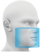
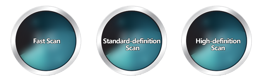
Option 1
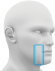
Option 2
Option 3
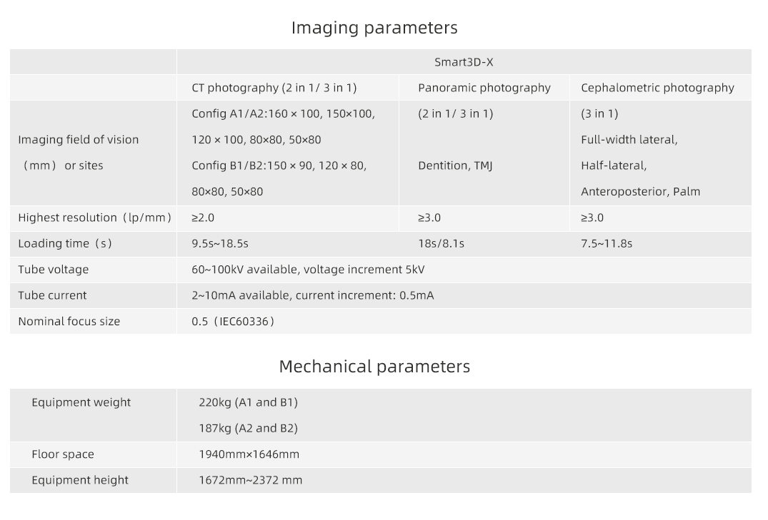
12x10cm
8x8cm
5x8cm
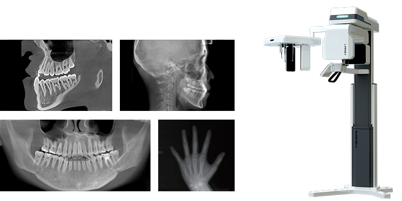
0.5mm small focus tube ensures outstanding image quality.
Resolution up to 2.0lp/mm, voxel size of 0.25~0.05 mm optional.

360°scan and 800 frame images with unique CT algorithms

QuartZ 4 scan platform, supporting flexible scan mode

Multiple focus layers in panoramic imaging, fitting the patient's dental arch

Easy-to-target scan area

Six positioning lasers with face-to-face communication to posit precisely

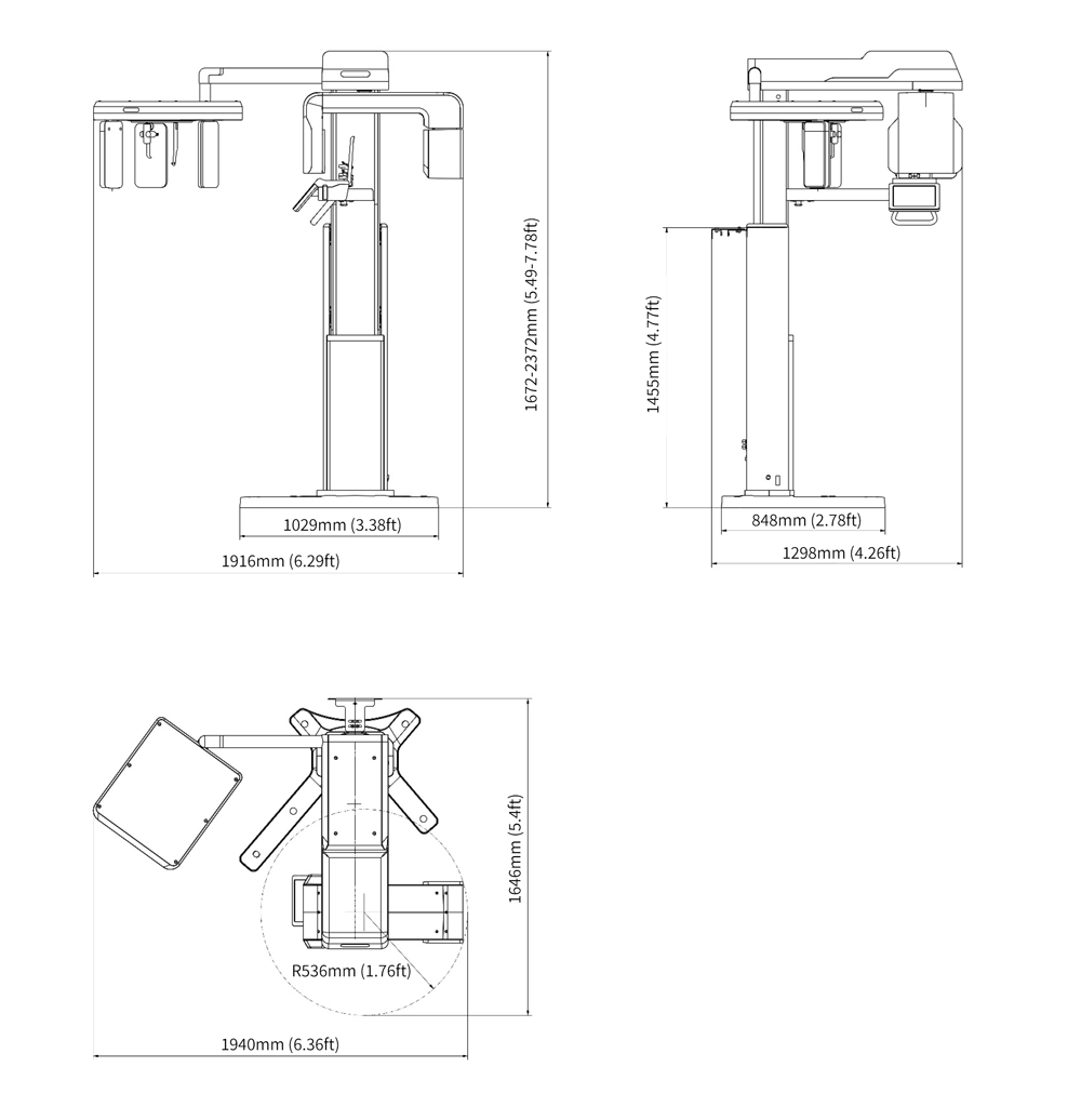
X-type base is convenient for wheelchair-bound patients

10"LED touch screen

Storage box design

Voice reminder
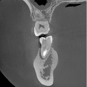
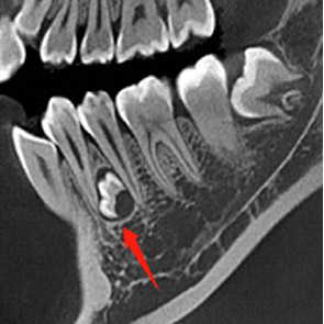
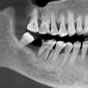
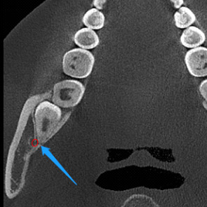
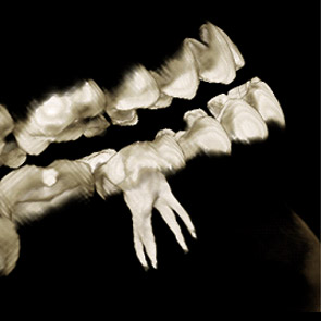
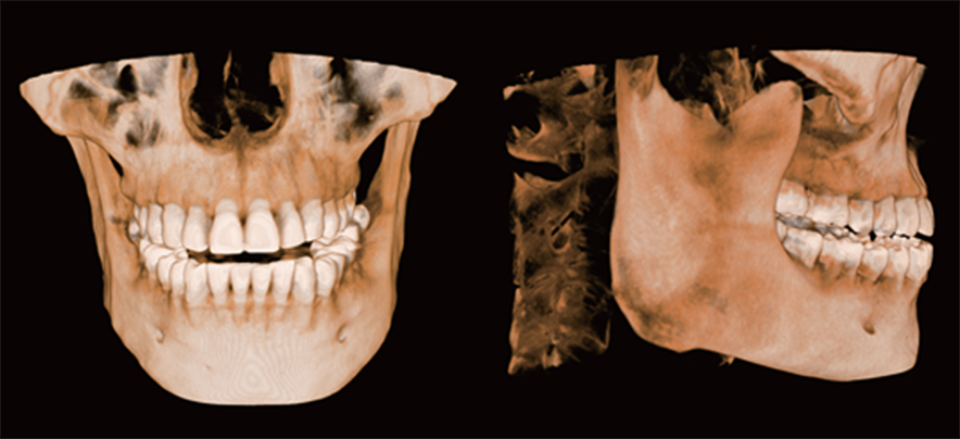
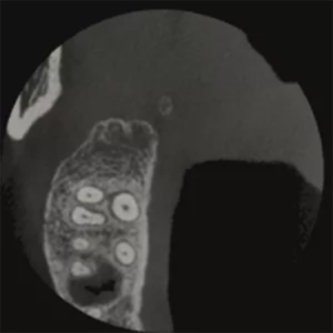
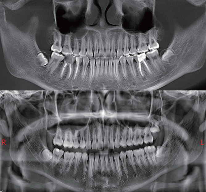
Axial, coronal and sagittal slices can be observed simultaneously. Besides, the slice in any direction is available. Buccolingual slices, distal and mesial sections were obtained to facilitate diagnosis.
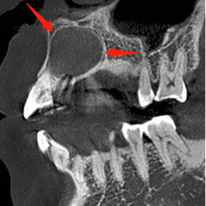
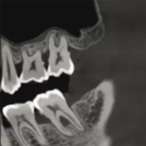
Local fine reconstruction is conducted in the designated area.
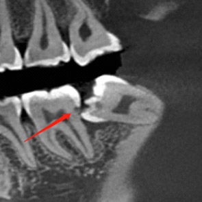
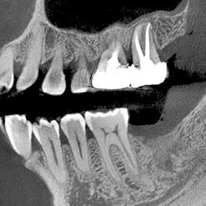
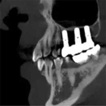
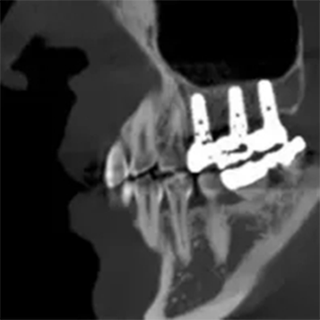
With the new T-MAR correction module for metal artifact removal, the system corrects metal artifacts intelligently. It avoids overmodification and saves the original clinical data.
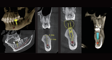
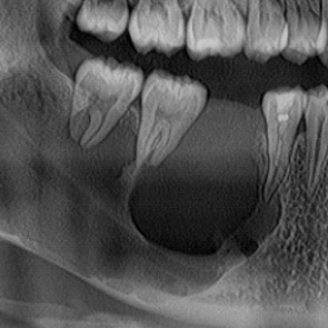
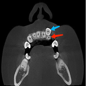
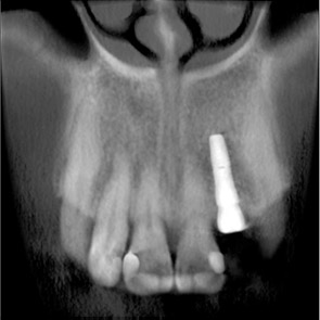
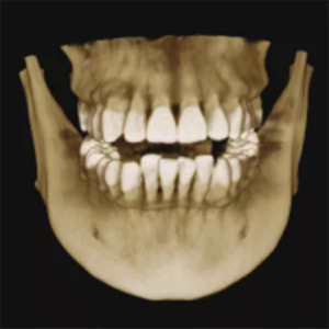
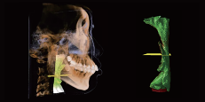
The bone and bone mass in the implant area will be evaluated by dental 3D images using HiRes3D. The neural tube will be highlighted automatically, which presents the relationship between the implant and the neural tube. This is a better way to approach a successful implant surgery.
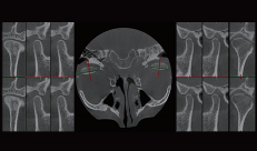
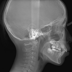
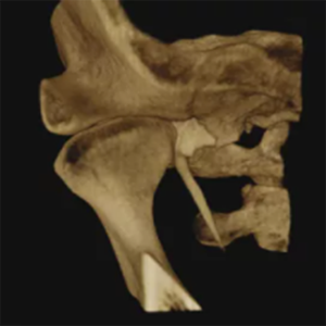
SmartVPro software has a visual pattern of comparing the left and right joints, allowing doctors to evaluate the diagnosis and treatment effect on temporomandibular joint diseases.
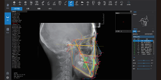
The neural network is trained by mega data, which automatically identifies orthodontic anatomical landmark points, draws anatomical structures and outputs measurement reports according to the selected measurement methods.
The airway is segmented automatically, which calculates the volume and the narrowest area of the airway.
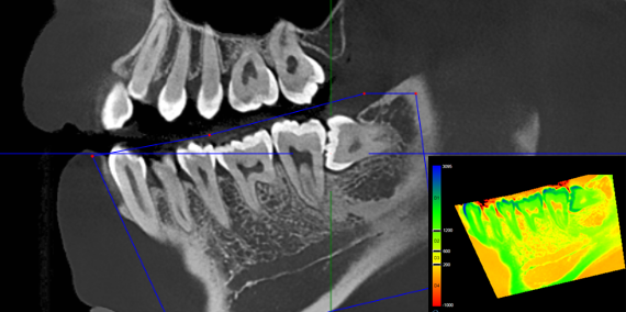
Used to assess bone mineral density in selected areas.